Pelvic Anatomy Female - Anatomy Of The Female Pelvis Springerlink / The greater pelvis is a part of the abdomen study section, we won't spend too long on that here.
Pelvic Anatomy Female - Anatomy Of The Female Pelvis Springerlink / The greater pelvis is a part of the abdomen study section, we won't spend too long on that here.. The lower third of the uterus comprises the cervix.the upper boundary of the cervix is the level of the internal os, a narrowing of the uterus that is also referred to as the isthmus.the internal os is the opening between the cervix and the corpus. Ct body (lymph nodes) ct. Anatomically correct, the language of the text is clear and concise. Über 7 millionen englischsprachige bücher. Female pelvis the female pelvis is broader and larger than the male pelvis to provide a comfortable environment for fetus development.
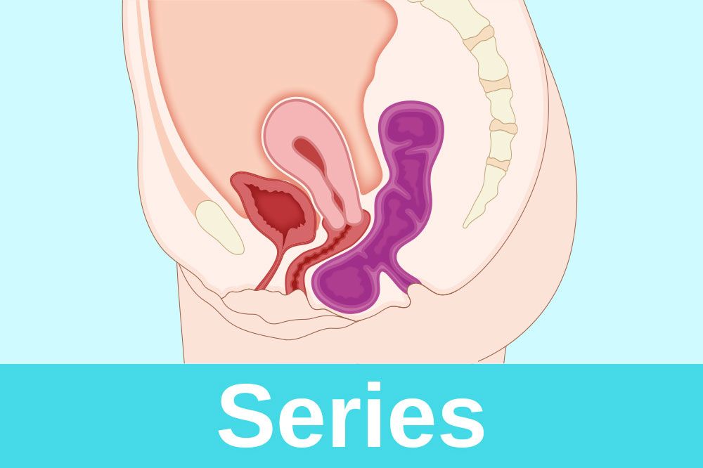
Structure and function the uterus sits in the center of the female pelvic cavity (figure 2.) the most common position of the uterus in the pelvic cavity is anteverted and anteflexed. This mri female pelvis axial cross sectional anatomy title tool is absolutely free to use. 4.3 out of 5 stars 27. The greater pelvis is a part of the abdomen study section, we won't spend too long on that here. Differentiate the different types of the female pelvis.
Female Pelvic Anatomy Model Teaching Of Gynecology And Obstetrics Medicine For Genitourinary Urinary Uterine Embryo And Fetus Educational Equipment Aliexpress from ae01.alicdn.com Anatomically correct, the language of the text is clear and concise. Describe the anatomy of the pelvic wall, bones, joints & muscles. A macroscopic study of the hypogastric plexus and their relations and variations j anat. In this image, you will find anatomy of female pelvic area in detail, suspensory ligament of ovary, paravesical pouch, broad ligament, mesovarium, ovary, uterine (fallopian) tube, round ligament of uterus, ligament of ovary, uterus, internal iliac artery and vein, linea terminalis, cervix, obturator membrane, obturator fascia in it. It's located between the abdomen and the legs. • divided into the true and false pelvis by the iliopectineal line. This video is a great representation of the work done by elara systems. They act to support the female viscera and provide a conduit for neurovascular structures.
Describe the anatomy of the pelvic wall, bones, joints & muscles.
Anatomy of the female pelvis. Surgical anatomy of the female pelvis by laparoscopy. • pelvis begins at the iliac crests and ends at the symphysis pubis. Describe the components & function of the pelvic diaphragm. Über 7 millionen englischsprachige bücher. I'm so glad you're joining me as i take you on a user friendly and useful guide to understanding the female pelvis, her relationships and how to assess her! The greater pelvis is a part of the abdomen study section, we won't spend too long on that here. The shape of the female bony pelvis can be classified into four broad categories: This video is a great representation of the work done by elara systems. The pelvis's frame is made up of the bones of the pelvis, which connect the axial skeleton to the femurs, and therefore acts in weight bearing of the upper body. In this medical 3d animation, we take a look at the female pelvis anatomy. Gynecoid, anthropoid, android, and platypelloid 2. List the arterial & nerve supply list the lymph & venous drainage of the pelvis.
The pelvis's frame is made up of the bones of the pelvis, which connect the axial skeleton to the femurs, and therefore acts in weight bearing of the upper body. Laparoscopic anatomy of the female pelvic region. The sagittal (longitudinal) image of the female pelvis shows anatomical structures. Anatomically correct, the language of the text is clear and concise. The pelvis is the lower portion of the trunk, located between the abdomen and the lower limbs.
Female Pelvis Section Anatomy Model Used Within The Consultation Download Scientific Diagram from www.researchgate.net Gynecoid, anthropoid, android, and platypelloid 2. Get it as soon as wed, may 26. A macroscopic study of the hypogastric plexus and their relations and variations j anat. Anatomy of the female pelvic nerves: This mri female pelvis axial cross sectional anatomy title tool is absolutely free to use. In female it is adapted for child bearing. Describe the boundaries and subdivisions of the pelvis. Hi and welcome to my functional female pelvic anatomy course!
The bony pelvis in normal standing posture transmits the body weight of head, trunk and the upper extremities to the lower extremities.
Hi and welcome to my functional female pelvic anatomy course! Describe the anatomy of the pelvic wall, bones, joints & muscles. Anatomy of the female pelvic nerves: Ct body (lymph nodes) ct. Anatomy of the female pelvis. Gynecoid, anthropoid, android, and platypelloid 2. Laparoscopic anatomy of the female pelvic region. The term pelvic floor refers to all of the supportive structures that are involved with pelvic organ support. I'm so glad you're joining me as i take you on a user friendly and useful guide to understanding the female pelvis, her relationships and how to assess her! The shape of the female bony pelvis can be classified into four broad categories: Structure and function the uterus sits in the center of the female pelvic cavity (figure 2.) the most common position of the uterus in the pelvic cavity is anteverted and anteflexed. Use the mouse scroll wheel to move the images up and down alternatively use the tiny arrows (>>) on both side of the image to move the images.>>) on both side of the image to move the images. The female true pelvis differs from the male in being shallower, having straighter sides, a wider angle between the pubic rami at the symphysis, and a proportionately larger pelvic outlet.
Female pelvis the female pelvis is broader and larger than the male pelvis to provide a comfortable environment for fetus development. Just note that the terminal ileum, cecum and sigmoid colon are found within the greater pelvis in both sexes. Anatomically correct, the language of the text is clear and concise. Male and female, from in front and above, 1923. Laparoscopic anatomy of the female pelvic region.
Female Pelvic Floor 1 Anatomy And Pathophysiology Nursing Times from cdn.ps.emap.com The greater pelvis is a part of the abdomen study section, we won't spend too long on that here. 4.3 out of 5 stars 27. The obstetrical anatomy of a typical female pelvis is best considered as one unit. This mri female pelvis axial cross sectional anatomy title tool is absolutely free to use. Surgical anatomy of the female pelvis by laparoscopy. The pelvis is the lower portion of the trunk, located between the abdomen and the lower limbs. The bony pelvis in normal standing posture transmits the body weight of head, trunk and the upper extremities to the lower extremities. • pelvis begins at the iliac crests and ends at the symphysis pubis.
Male and female, from in front and above, 1923.
List the arterial & nerve supply list the lymph & venous drainage of the pelvis. Male and female, from in front and above, 1923. The lower third of the uterus comprises the cervix.the upper boundary of the cervix is the level of the internal os, a narrowing of the uterus that is also referred to as the isthmus.the internal os is the opening between the cervix and the corpus. Over 250 drawings illustrate every important aspect of pelvic anatomy, and show the reader how to perform simple exercises to keep the pelvis and its related structures fit. True pelvis • also known as pelvic cavity. This video is a great representation of the work done by elara systems. In this image, you will find anatomy of female pelvic area in detail, suspensory ligament of ovary, paravesical pouch, broad ligament, mesovarium, ovary, uterine (fallopian) tube, round ligament of uterus, ligament of ovary, uterus, internal iliac artery and vein, linea terminalis, cervix, obturator membrane, obturator fascia in it. In female it is adapted for child bearing. Pelvic diaphragm —the term pelvic diaphragm refers to the levator ani muscle and its covering fasciae, both the superior fascia and the inferior fascia. They also include the vagina, which is the entryway to the uterus. Structure and function the uterus sits in the center of the female pelvic cavity (figure 2.) the most common position of the uterus in the pelvic cavity is anteverted and anteflexed. Describe the anatomy of the pelvic wall, bones, joints & muscles. Über 7 millionen englischsprachige bücher.
43 out of 5 stars 27 pelvic anatomy. Use the mouse scroll wheel to move the images up and down alternatively use the tiny arrows (>>) on both side of the image to move the images.>>) on both side of the image to move the images.


0 Komentar